Chapter 25 of 50
Let There Be Bioluminescence
« Arithmétique! algèbre! géométrie! trinité grandiose!triangle lumineux! Celui qui ne vous a pas connues est un insensé! »
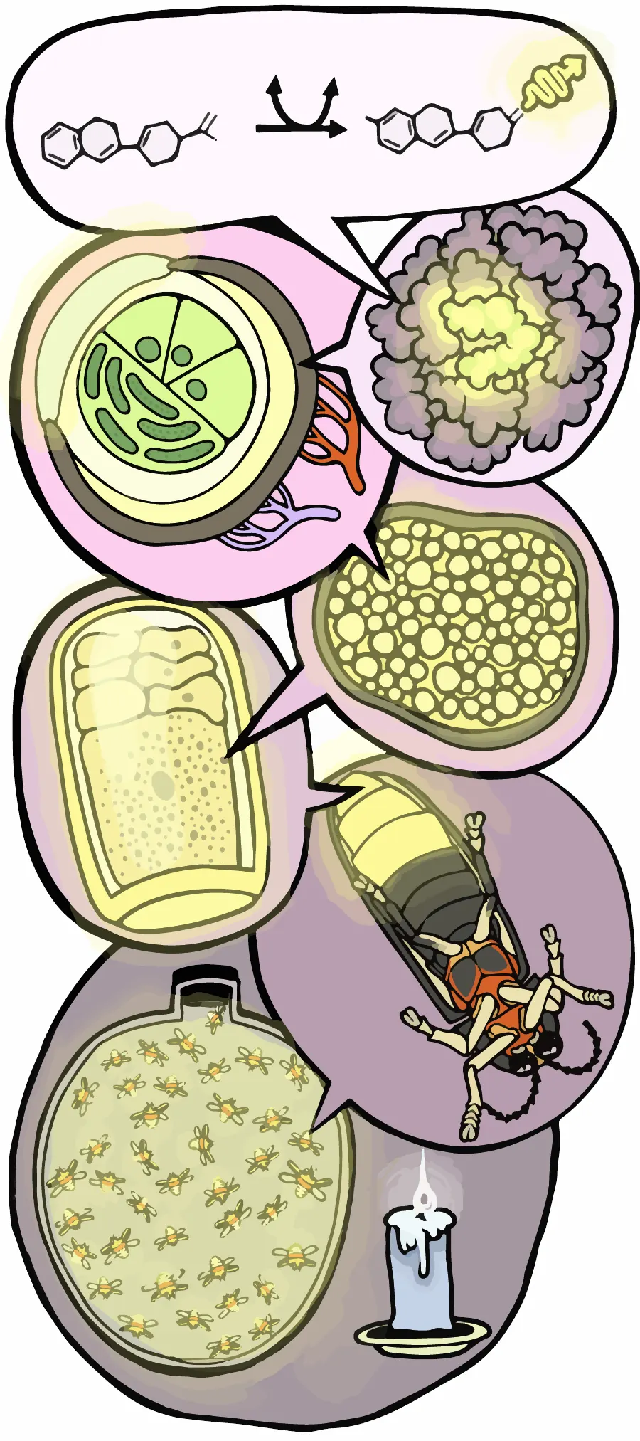
Firefly flashes demonstrate biology’s hierarchical organization from ecosystems to quantum mechanics. Species-specific flash patterns enable mate recognition and, in tropical swarms, synchronous displays visible across forests. Neural circuits generate these patterns by controlling oxygen flow through tracheal valves to specialized photocytes. Within these cells, luciferase catalyzes luciferin oxidation with extreme efficiency, converting chemical energy to light with minimal heat. The photons themselves arise when excited electrons in oxyluciferin transition between quantum energy states, emitting at 560–590 nm.
Topics
- Firefly Flash Patterns
- Luciferin-Luciferase Reaction
- Quantum Photon Emission
- Species-Specific Signals
- Flash Synchrony
- Photocyte Structure
- Oxygen Control
- 560nm Yellow-Green
- Photuris Mimicry
- Quantum Efficiency
- Transdisciplinary Cascade
Sidenote
Top (Molecular Ångström scale, reaction begins): At the Ångström scale, the luminescent reaction begins: luciferin is enzymatically converted into luciferyl-adenylate, then oxidized into oxyluciferin. The process releases a visible photon (), producing light.
Second (Enzyme Active Site): At nanometer scale, the luciferase enzyme forms a pocket where the reaction occurs. This active site holds luciferin in the correct orientation, enabling efficient light-producing catalysis.
Third (Subcellular Photocyte): At the 1–10 μm scale, we zoom into a photocyte — a specialized light-producing cell. Organelles such as mitochondria and endoplasmic reticulum support energy-demanding luminescence.
Fourth (Cell/Tissue Cross-Section): At the 10–100 μm scale, we see a full photocyte embedded in a patterned tissue. The densely packed spheres represent light-emitting units; organization optimizes light output and diffusion.
Fifth (Lantern Organ): At the 1–5 mm scale, the firefly’s lantern organ is visible in cross-section. Photocytes, tracheal tubes (for oxygen), and reflector layers are arranged to maximize brightness and directionality.
Sixth (Whole Firefly): At the centimeter scale, we see the full insect with its ventral lantern exposed.
Bottom (Population-Level Output): At the meter scale, we zoom out to an ecological view: a jar of approximately forty fireflies generates light that seems equivalent to a candle.